Because of a lapse in government funding, the information on this website may not be up to date, transactions
submitted via the website may not be processed, and the agency may not be able to respond to inquiries until
appropriations are enacted.
The NIH Clinical Center (the research hospital of NIH) is open. For more details about its operating status, please
visit cc.nih.gov.
Updates regarding government operating status and resumption of normal operations can be found at OPM.gov.
Ante la falta de fondos del gobierno federal, no se actualizará este sitio web y la organización no responderá
a transacciones ni consultas hasta que se aprueben los fondos.
El Centro Clínico de los Institutos Nacionales de la Salud (el hospital de investigación) permanecerá abierto.
Consulte cc.nih.gov (en
inglés).
Infórmese sobre el funcionamiento del gobierno federal y el reinicio de las actividades en
opm.gov.
This cow was inoculated with BPV1 virus by Carl Olson at the University of Wisconsin. The warts were harvested by Carl Baker and Paul Lambert and the material obtained (virus, tissue sections, RNA) was used for many of the key studies of BPV1 biology.
Author: Baker
Credit: Carl Baker and Paul Lambert. This image is the work of an National Institutes of Health employee, taken or made as part of that person's official duties. As a work of the U.S. federal government, the image is in the public domain.
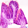
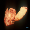
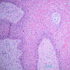
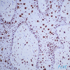
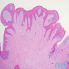
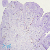
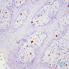
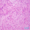
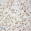
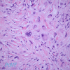
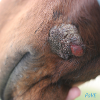
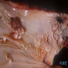
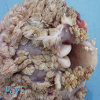
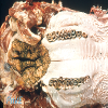
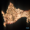
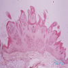
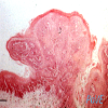
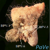
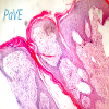
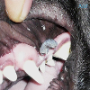
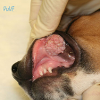
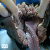
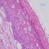
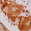
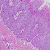
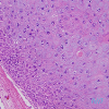
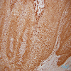
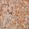
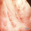
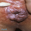
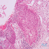
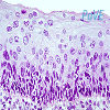
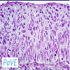
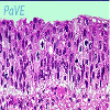
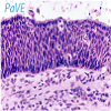
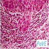
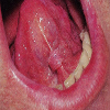
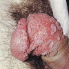
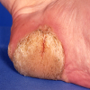
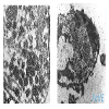
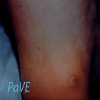
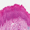
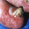
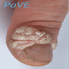
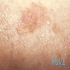
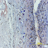
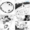
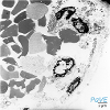
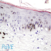
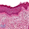
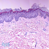
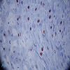
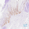
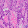
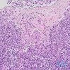
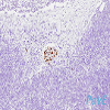
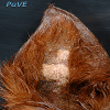
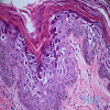
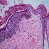
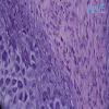
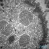
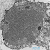
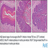
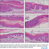
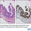
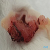
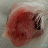
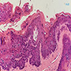
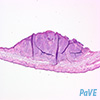
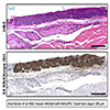
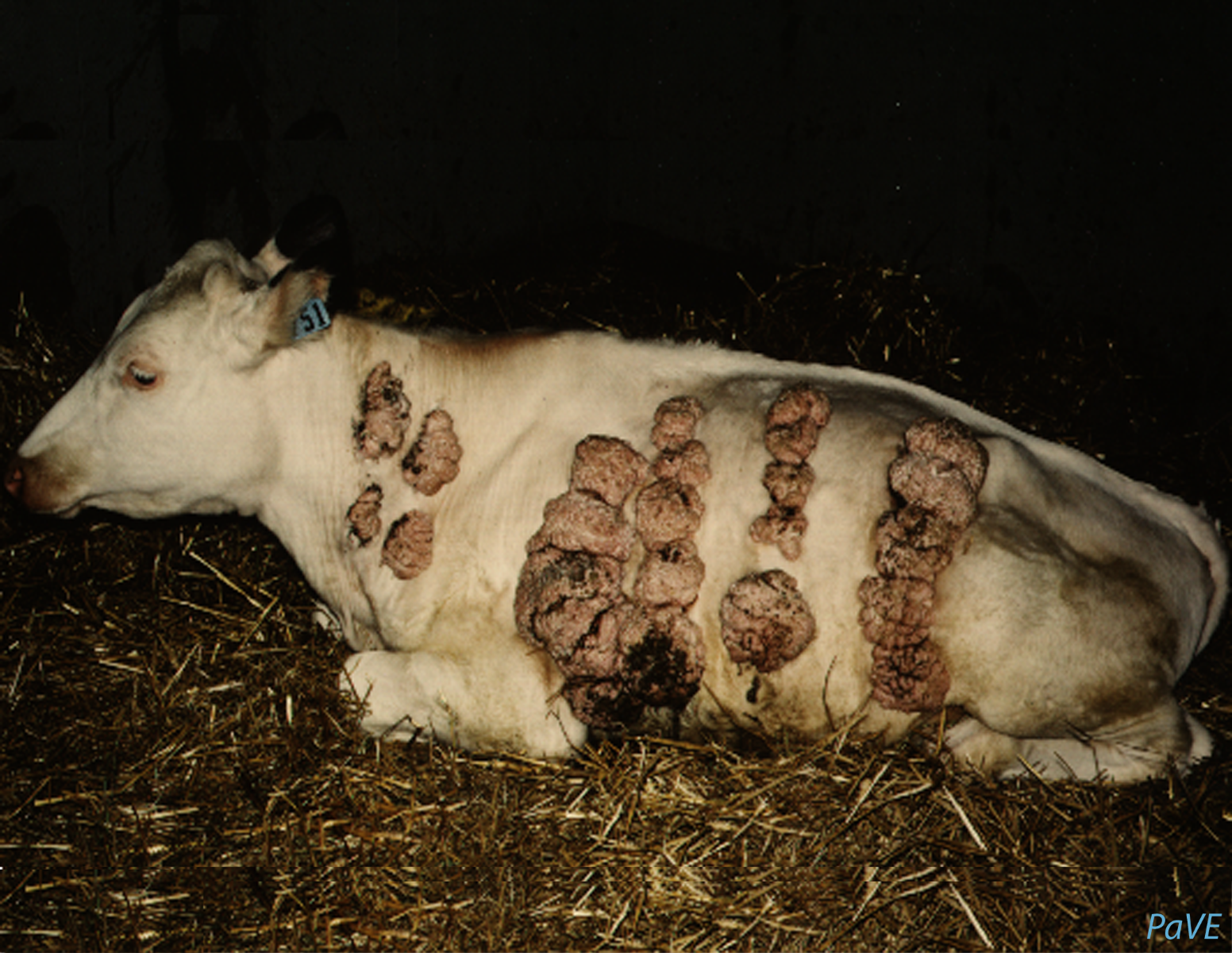
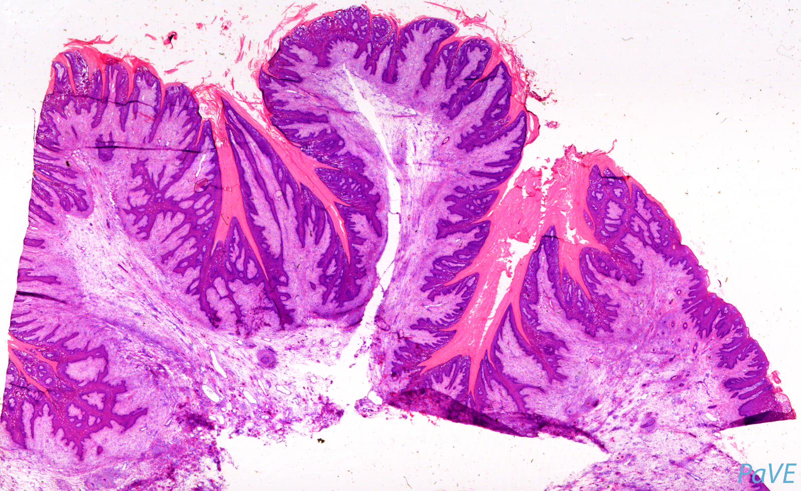
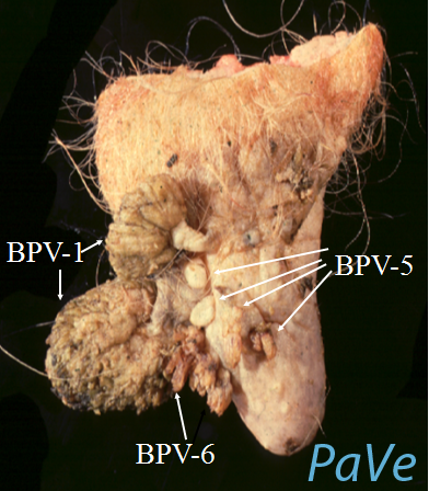
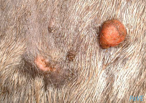
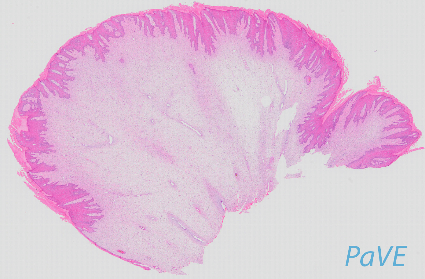
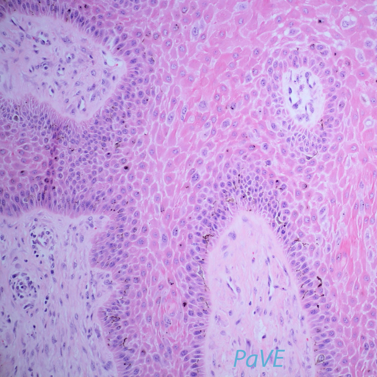
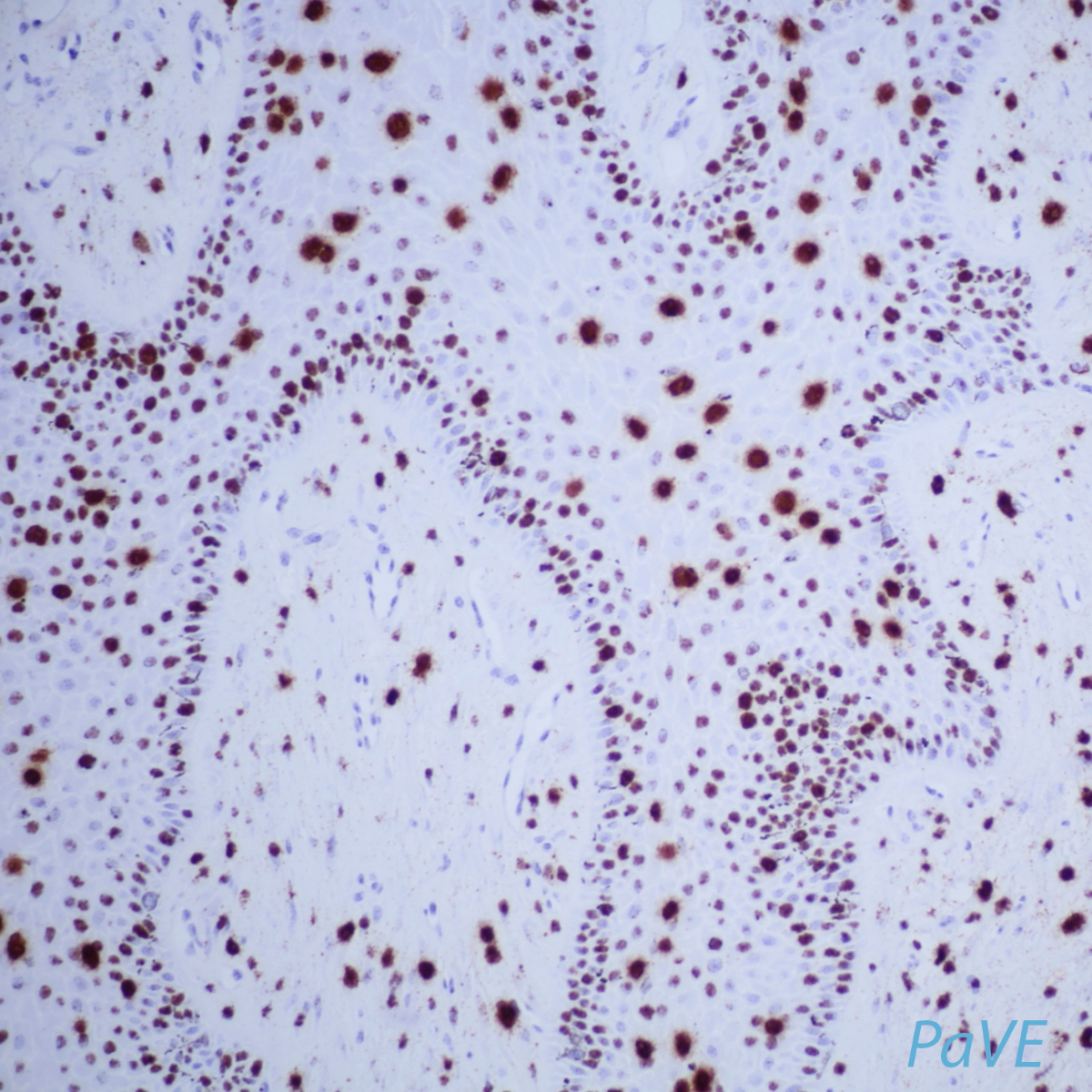
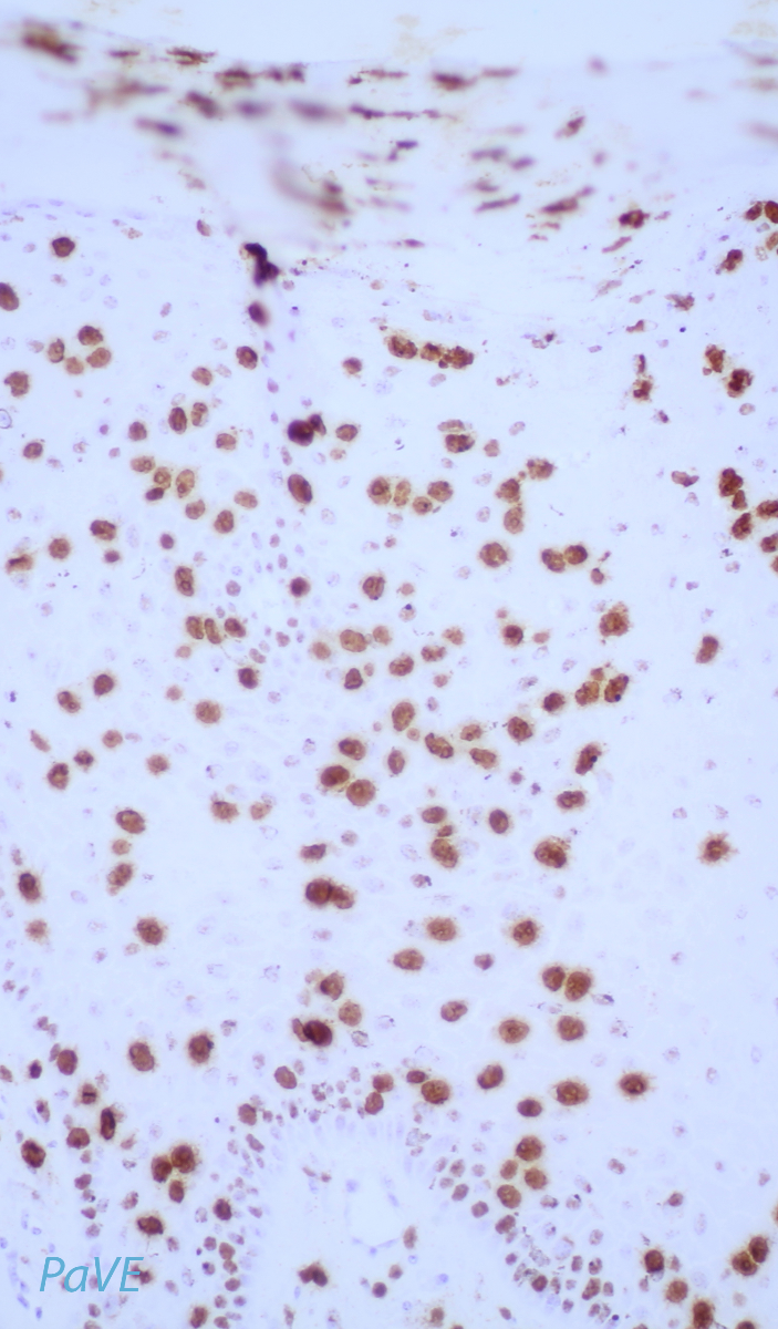
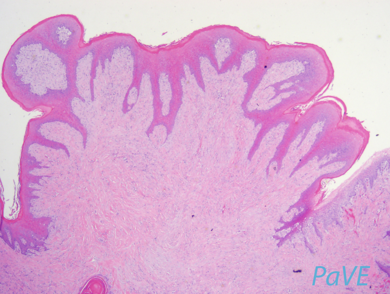
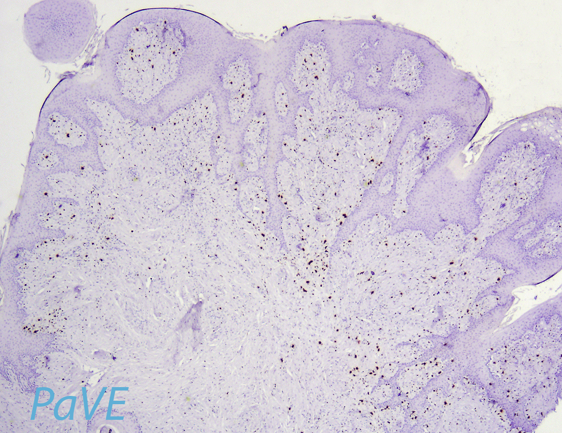
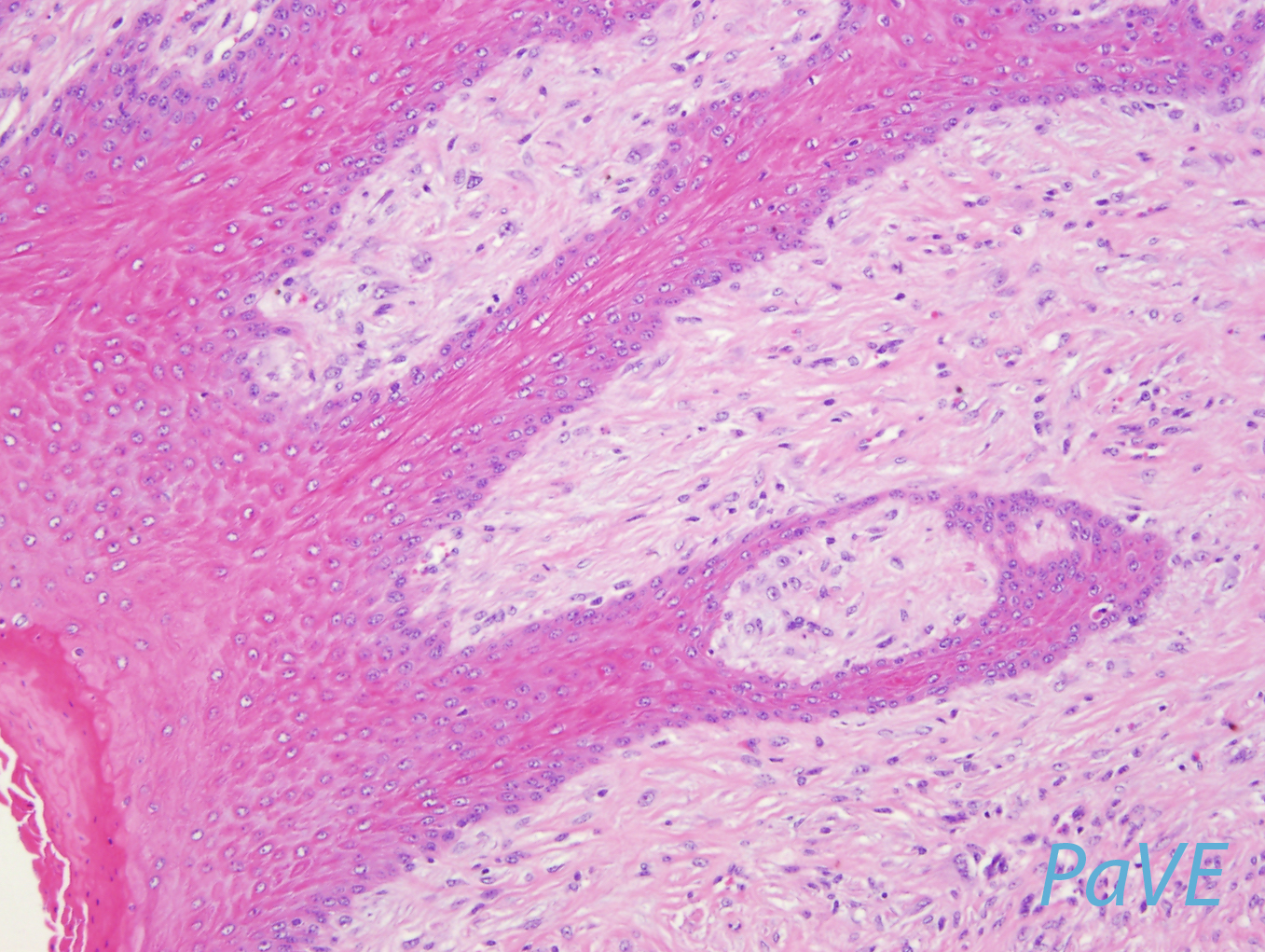
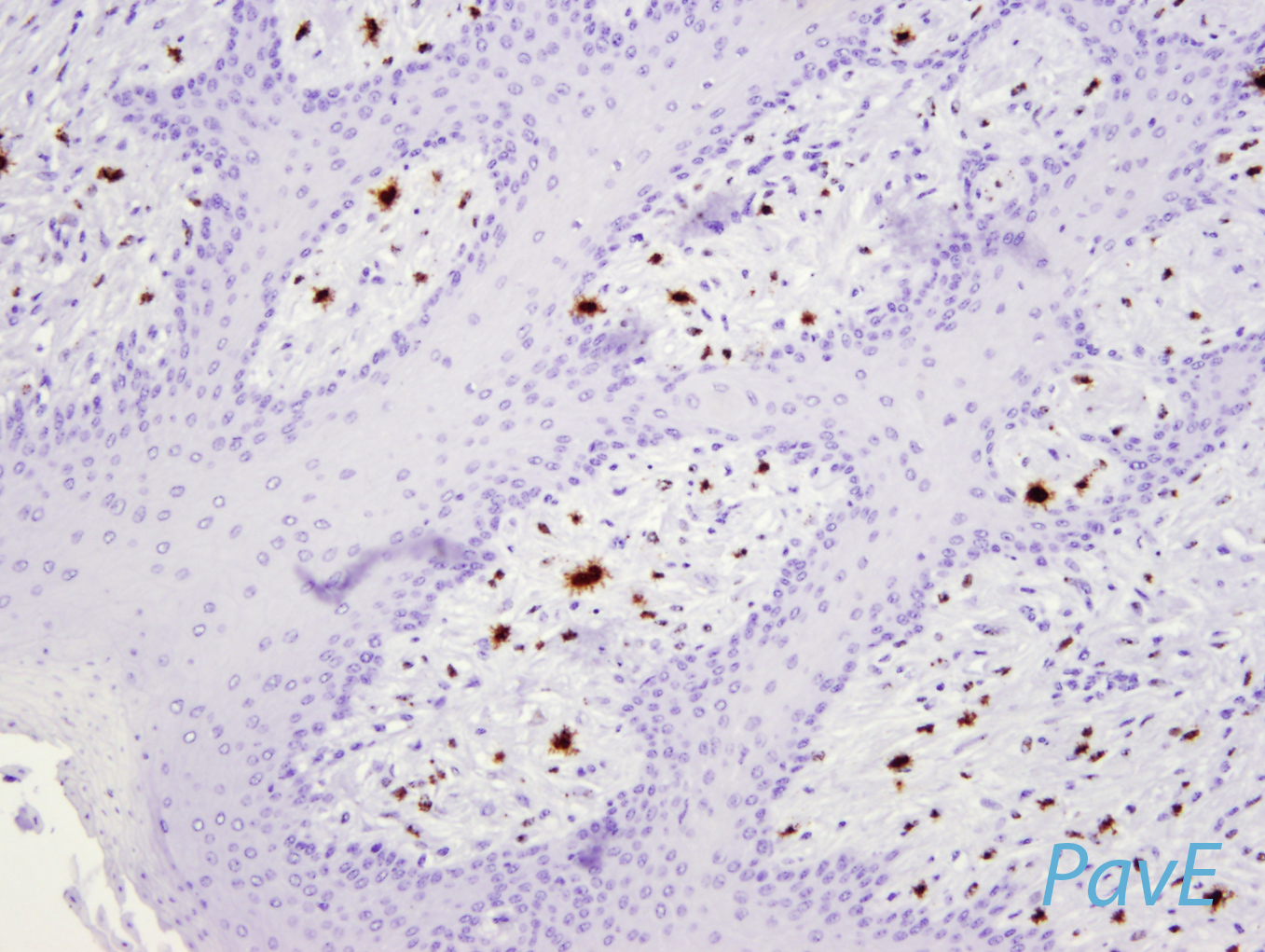
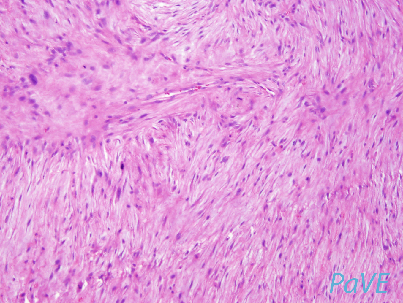
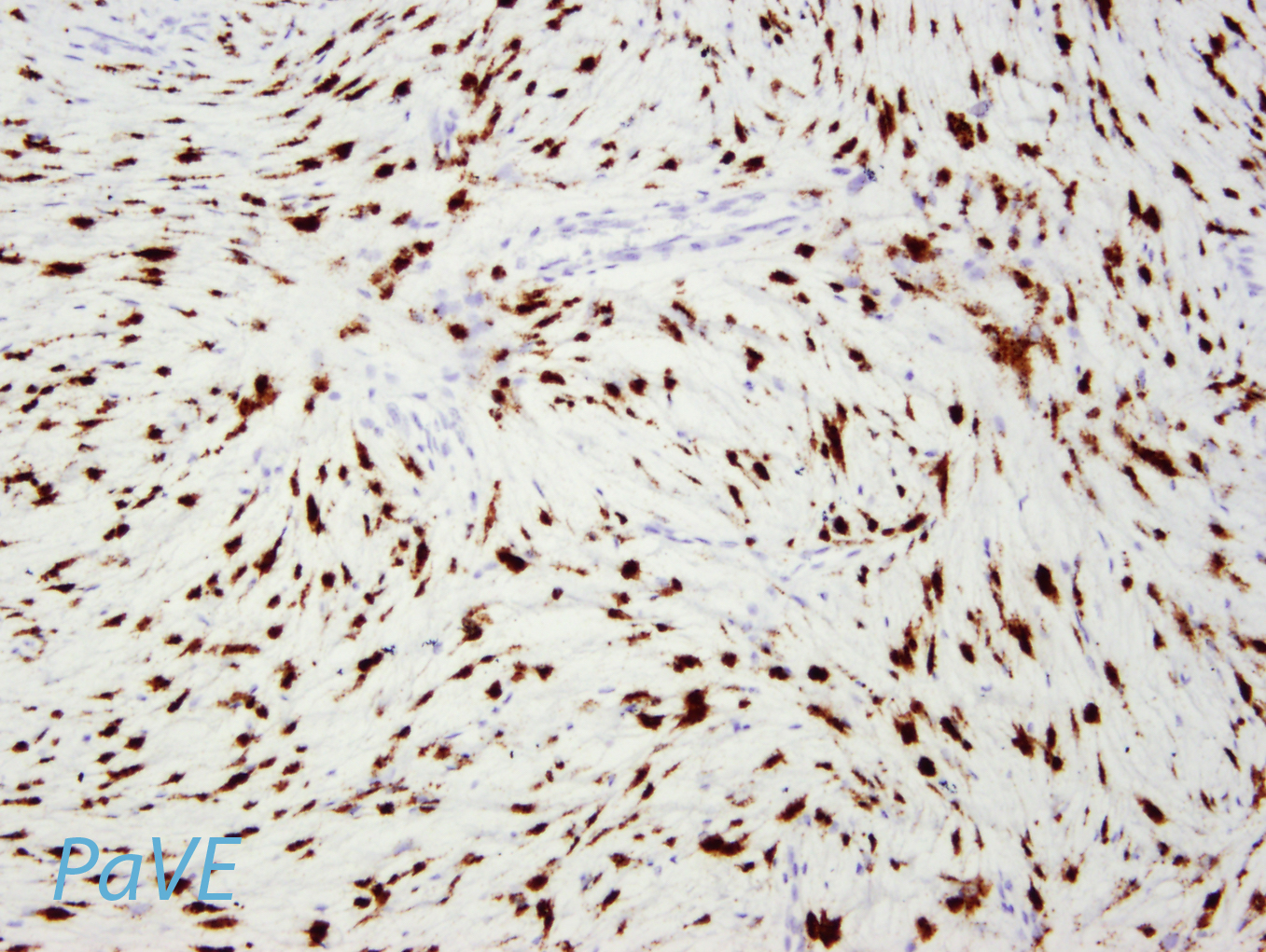
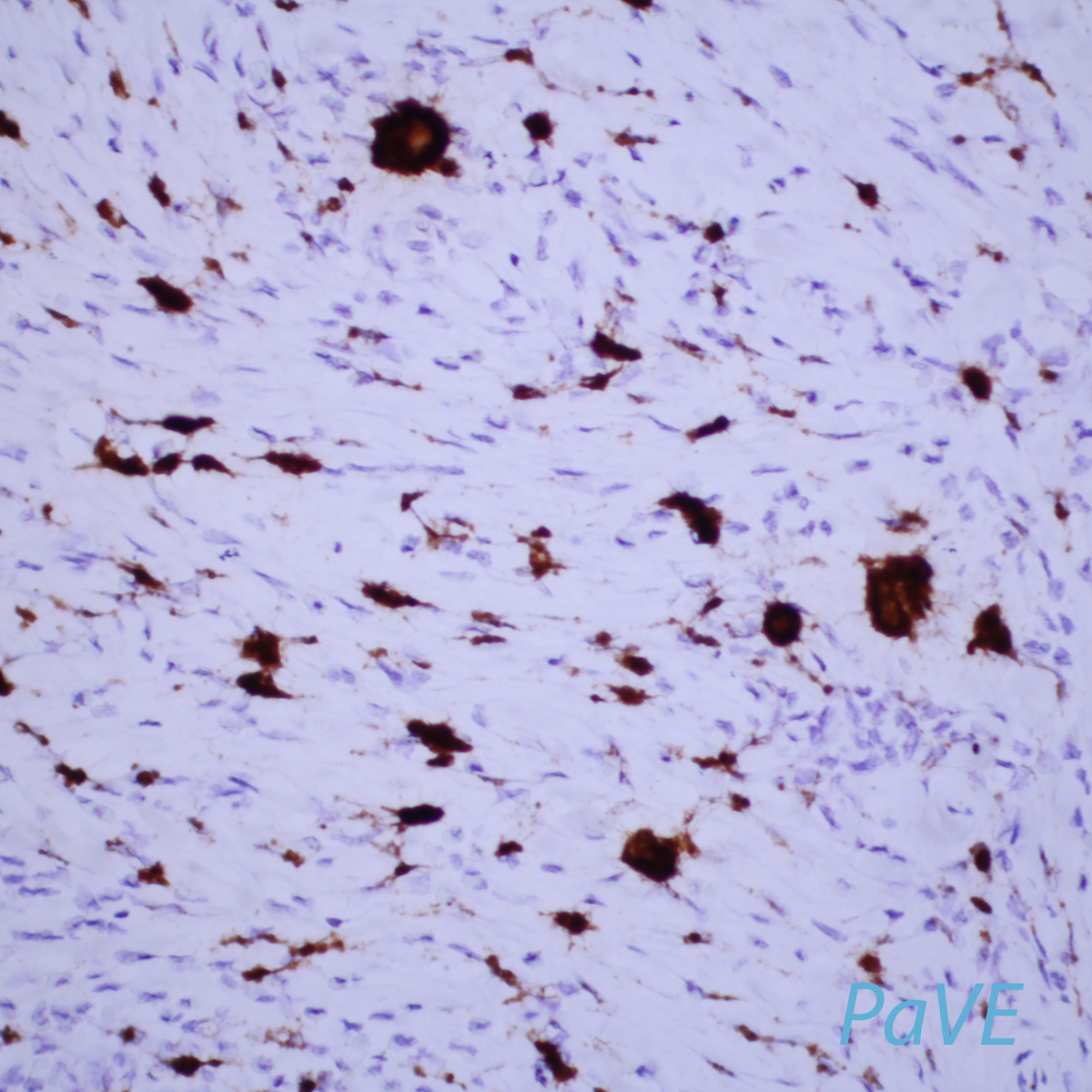
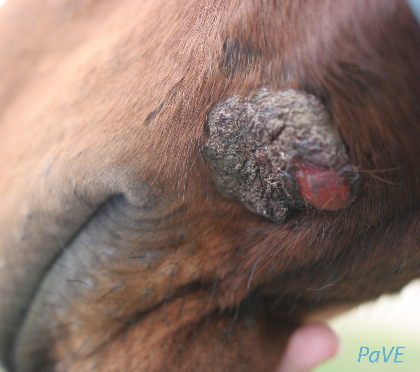
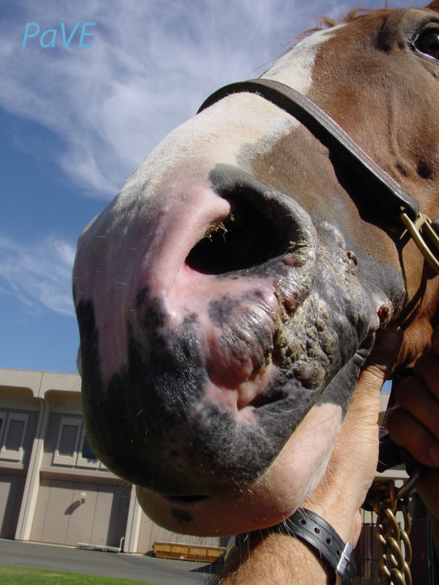
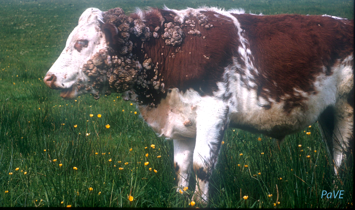
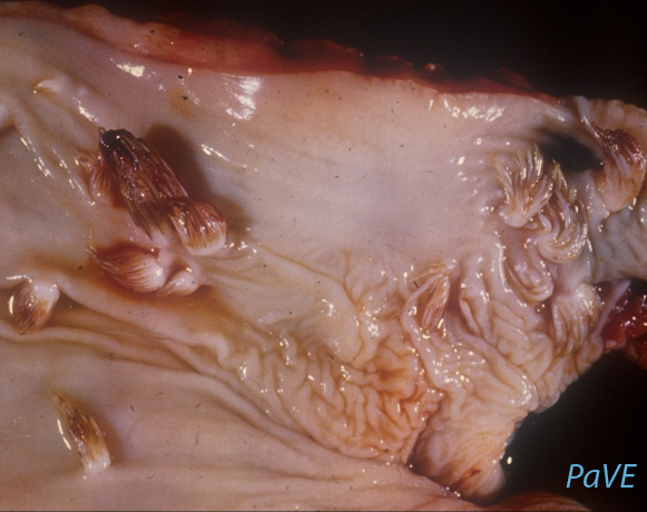
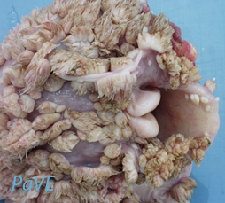
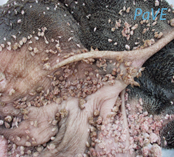
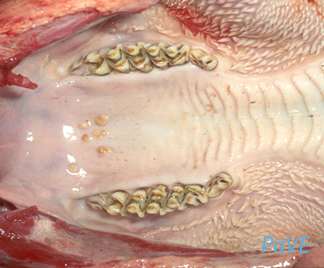
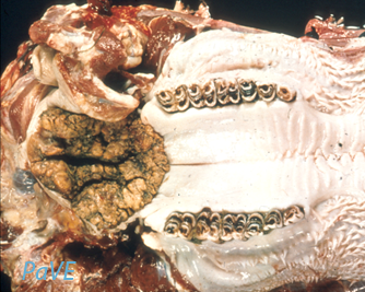
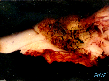
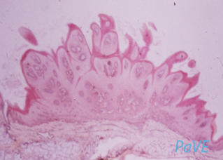
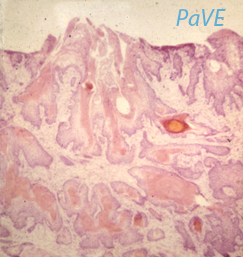
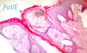
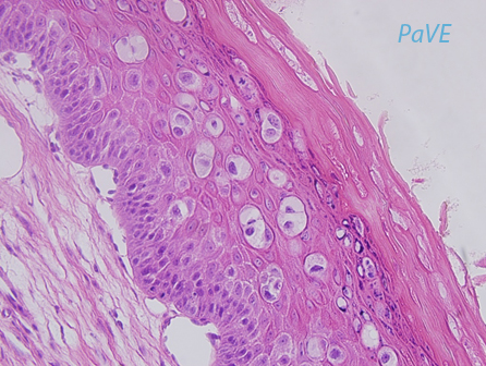
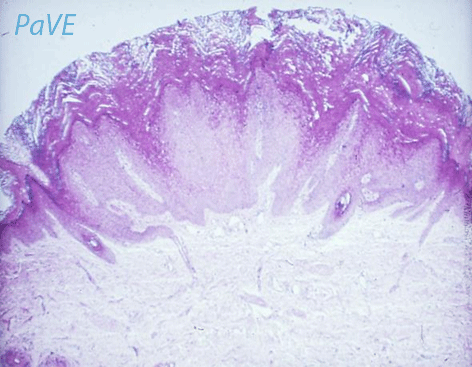
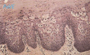
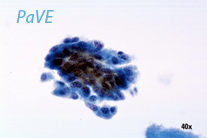
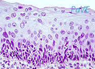
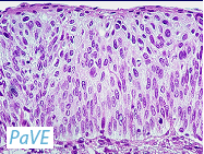
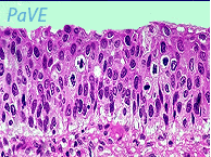
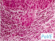
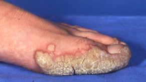
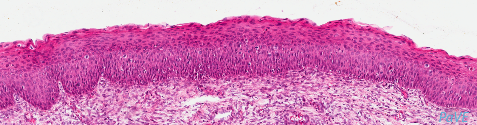
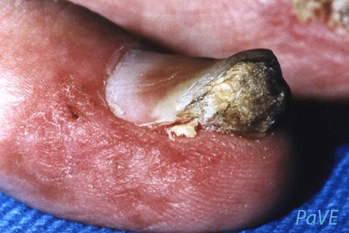
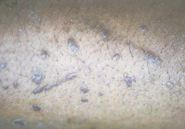
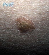
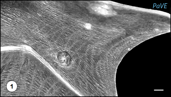
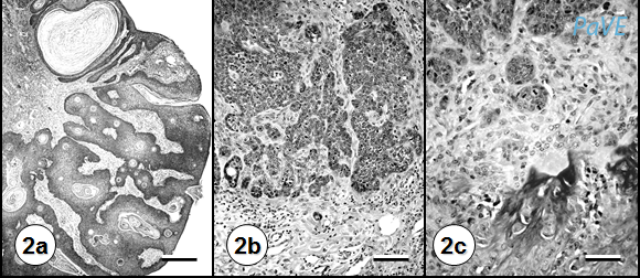
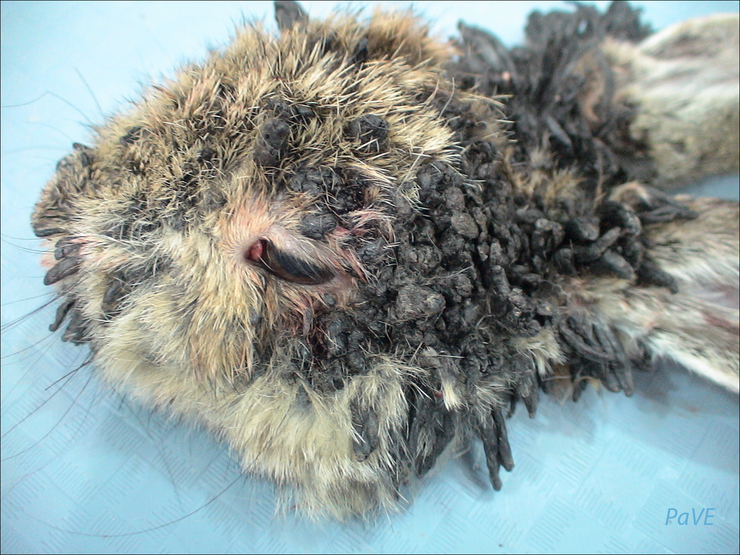
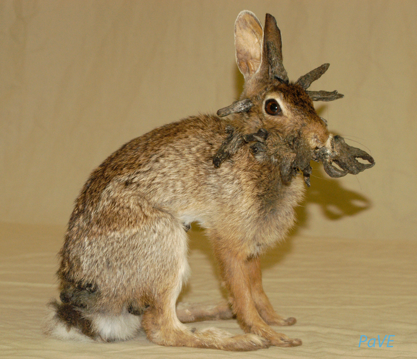
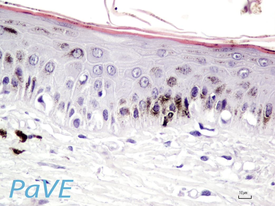
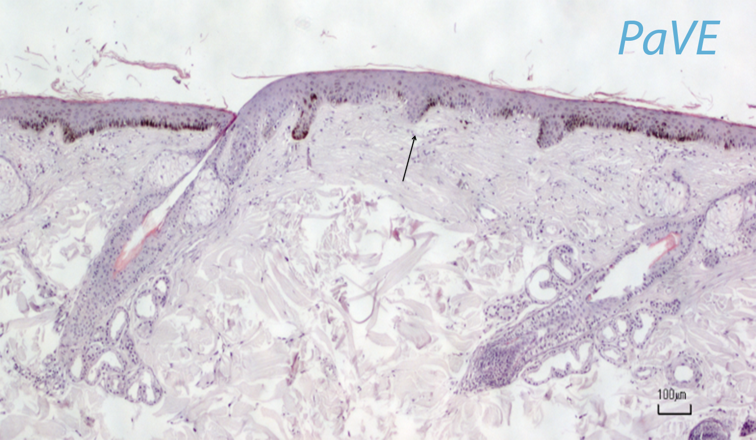
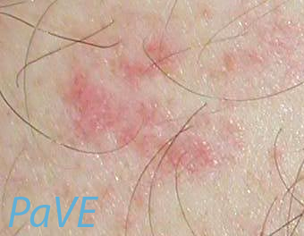
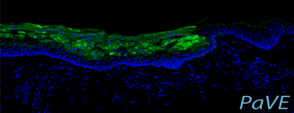
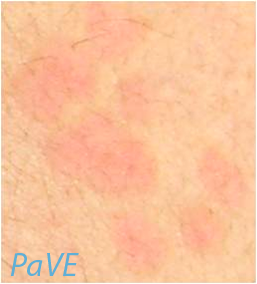
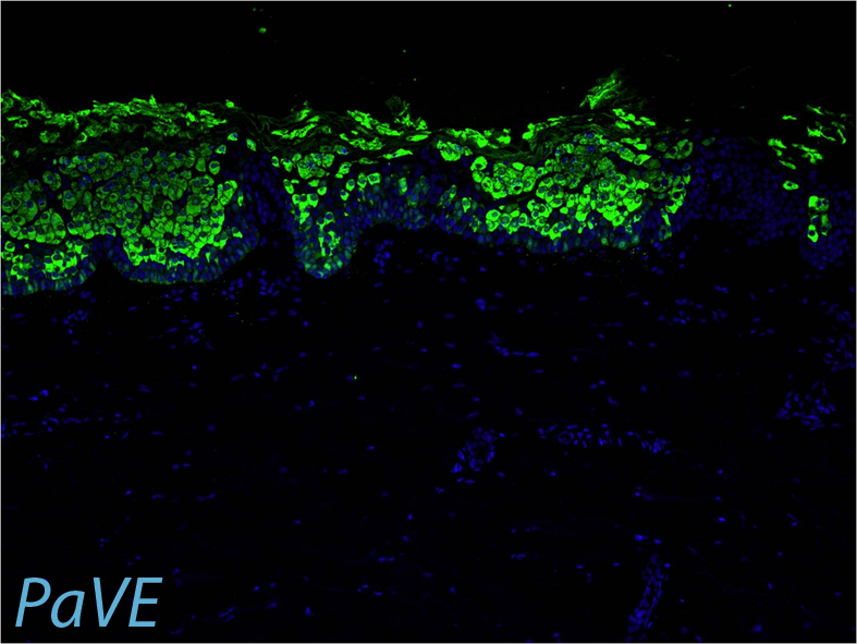
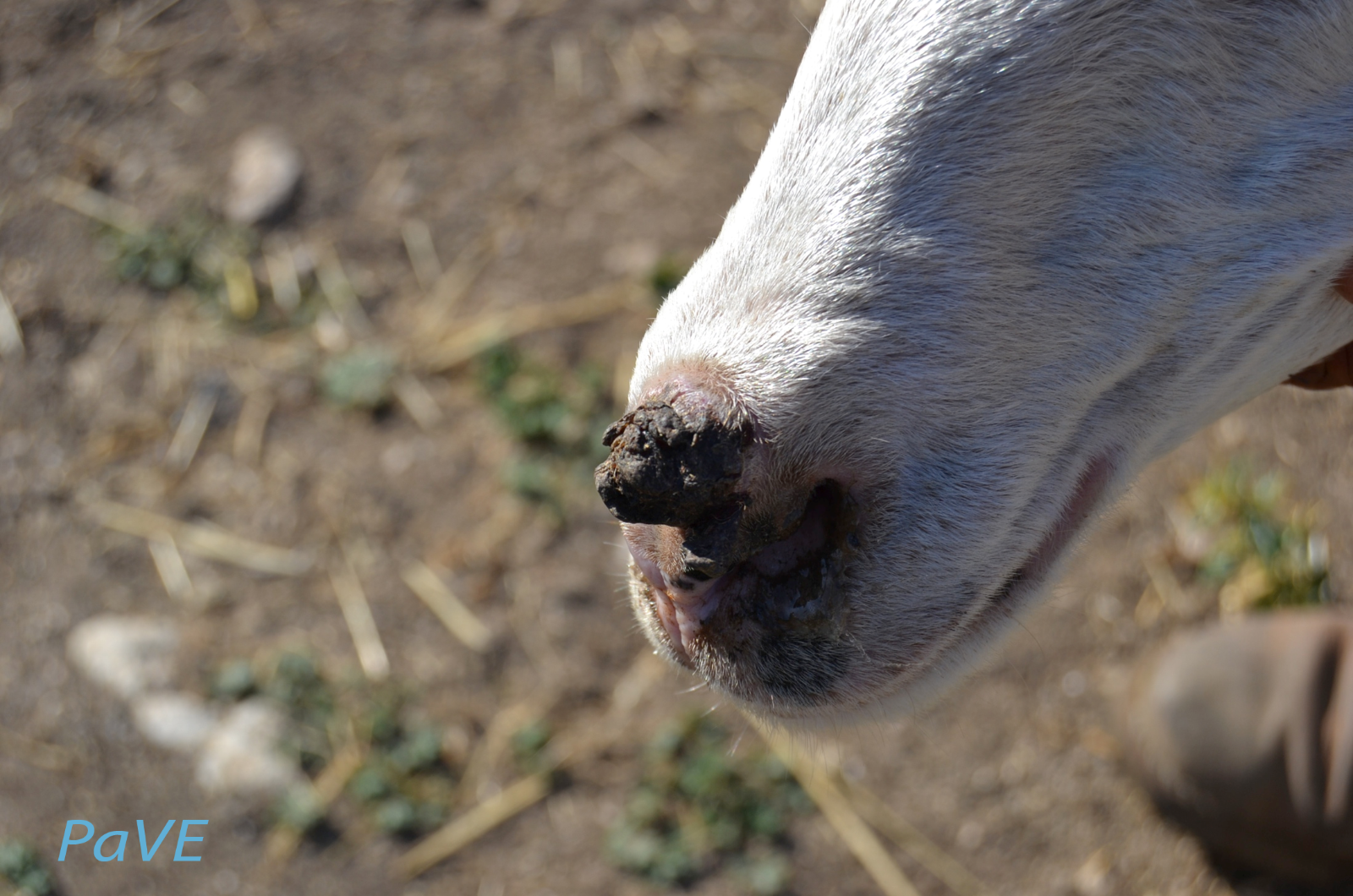
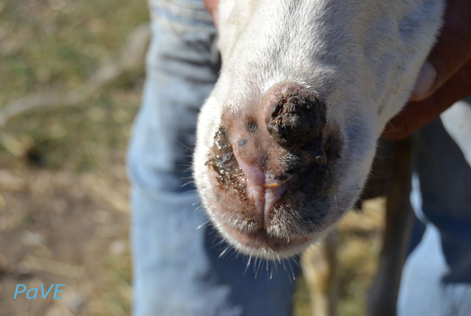
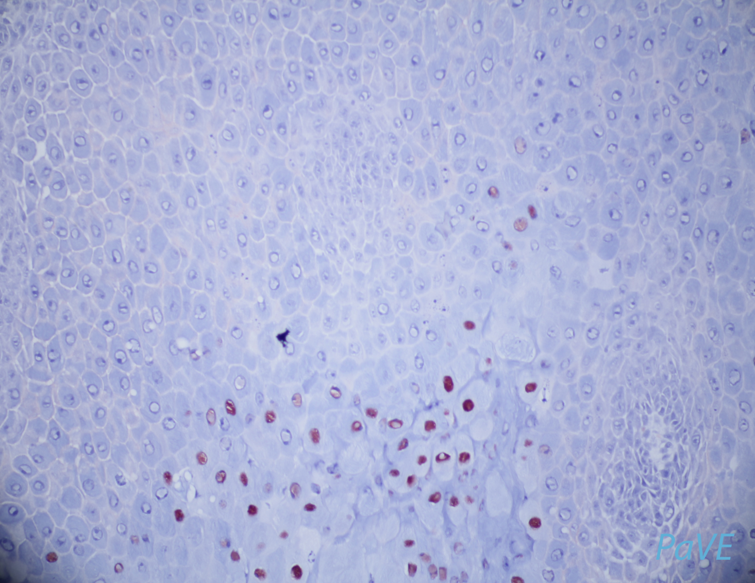
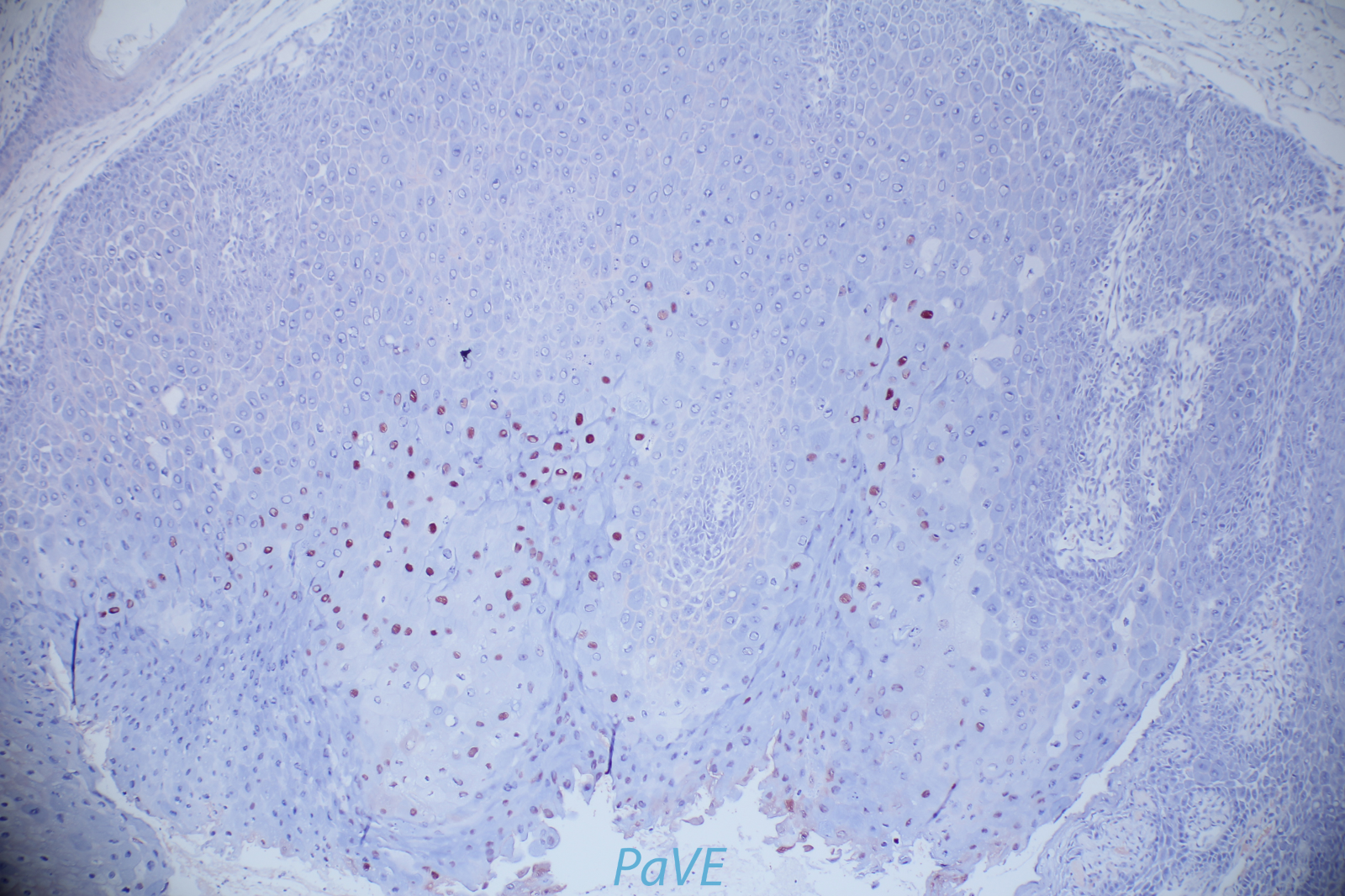
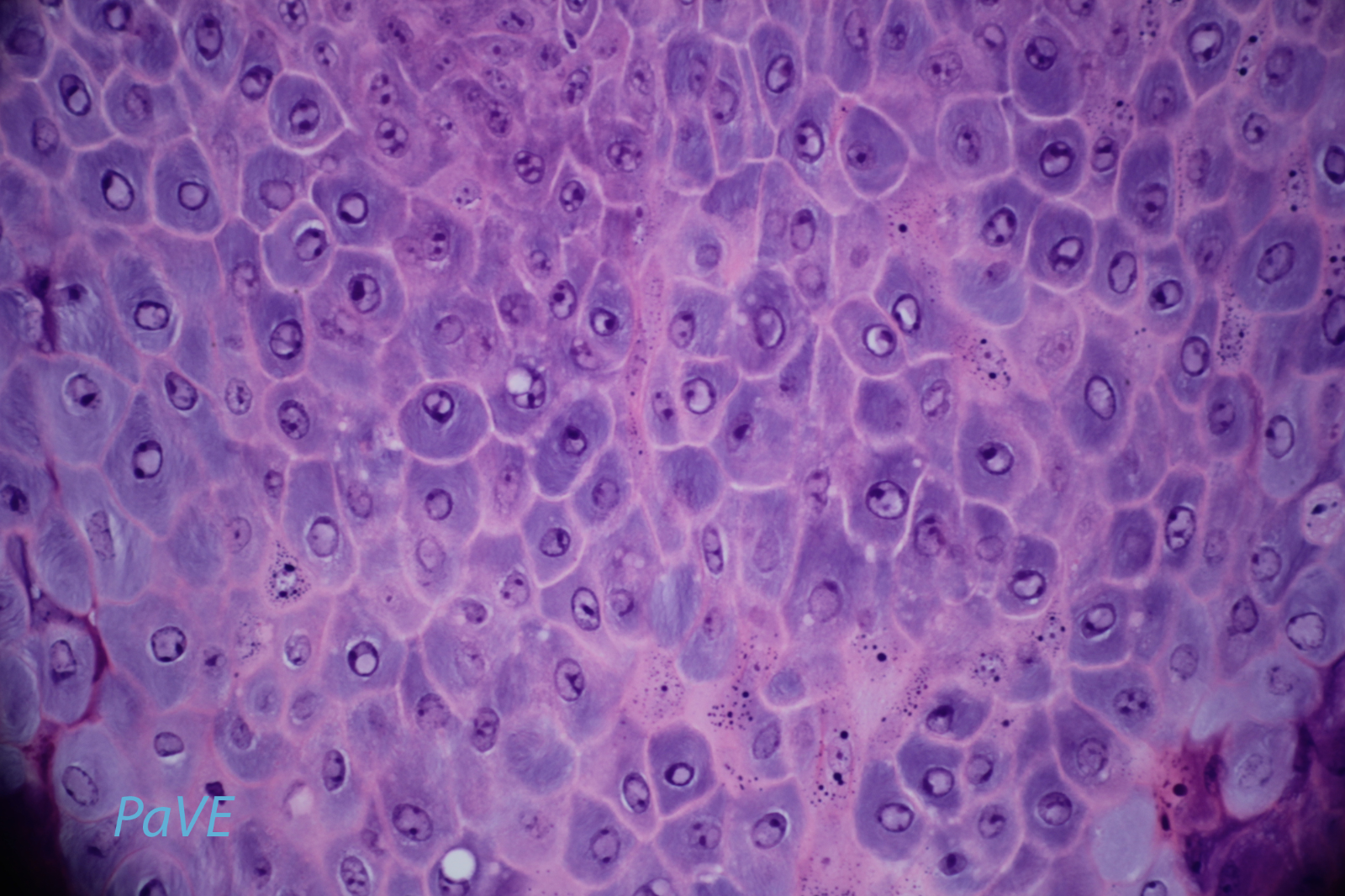
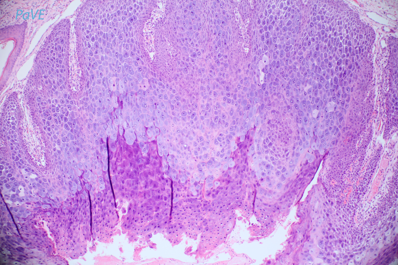
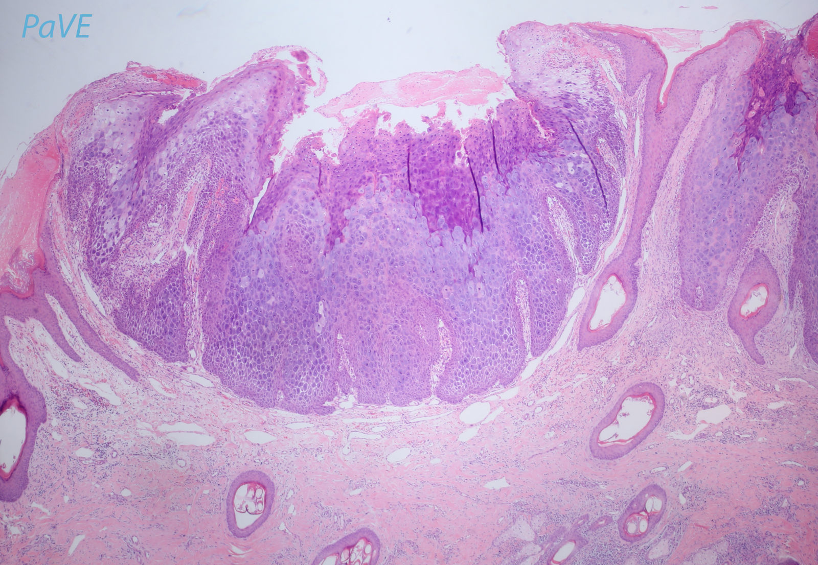
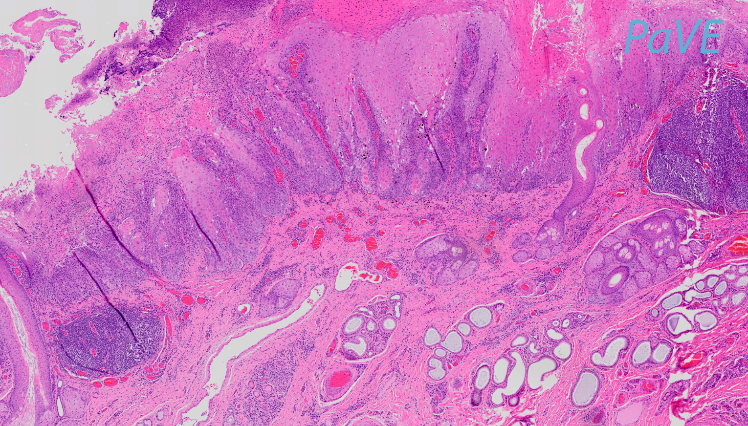
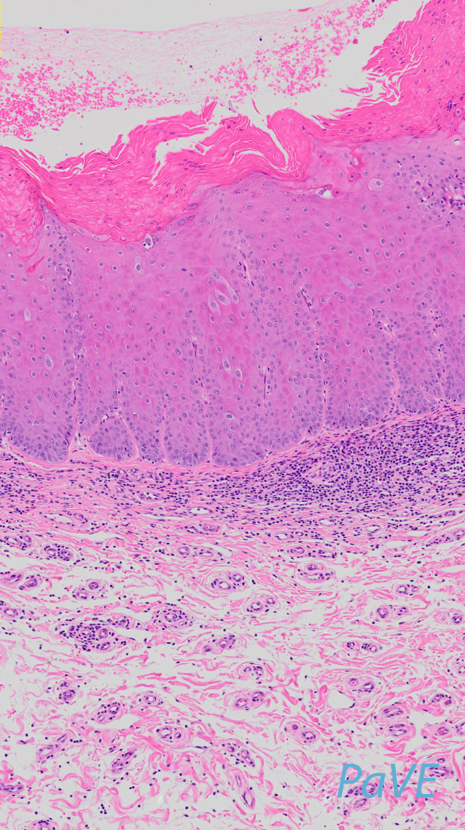
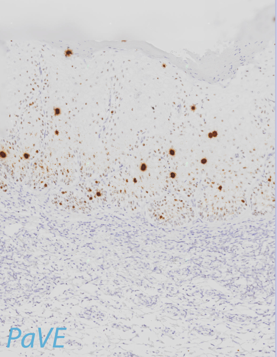
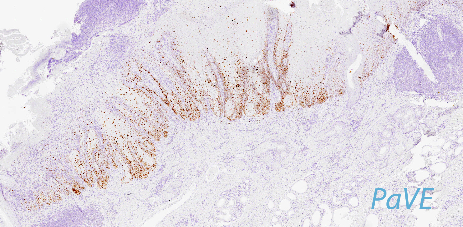
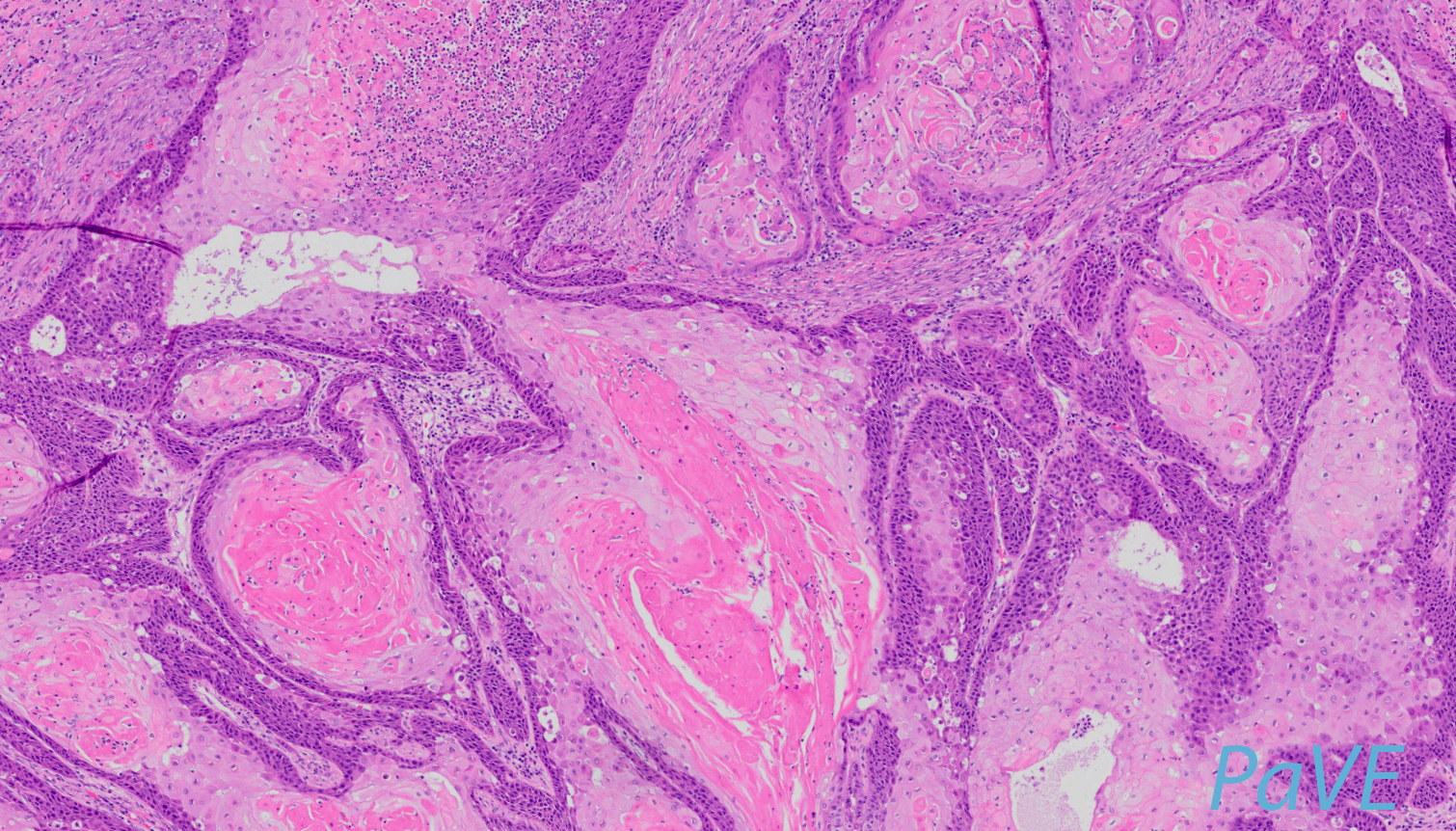
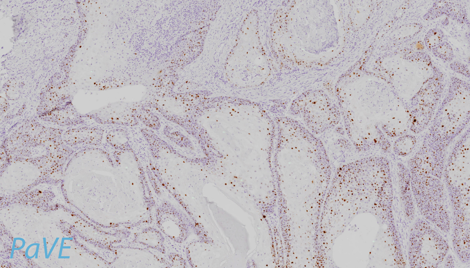
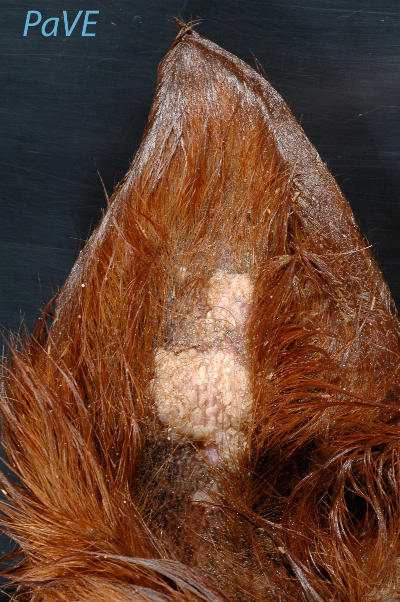
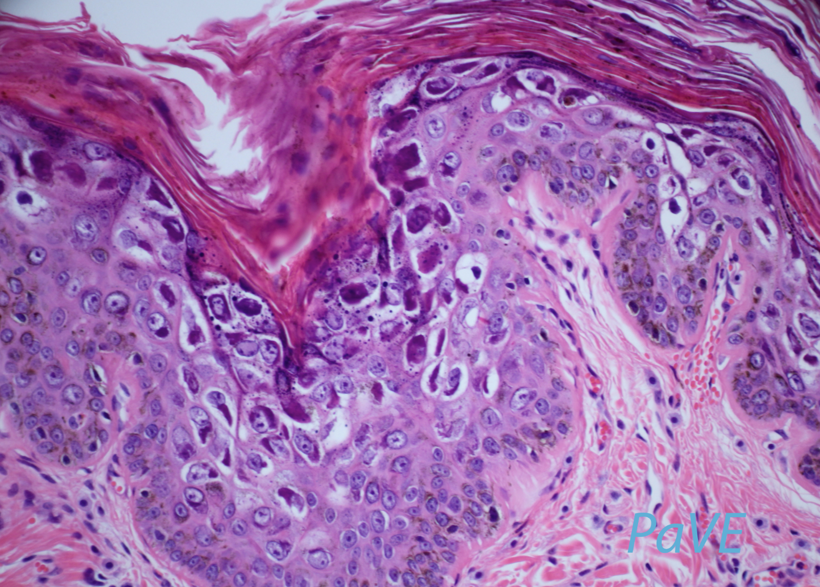
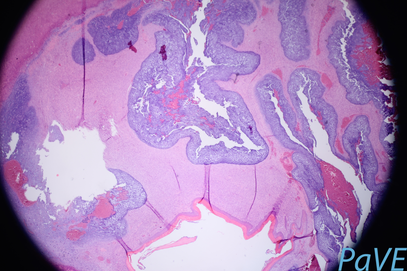
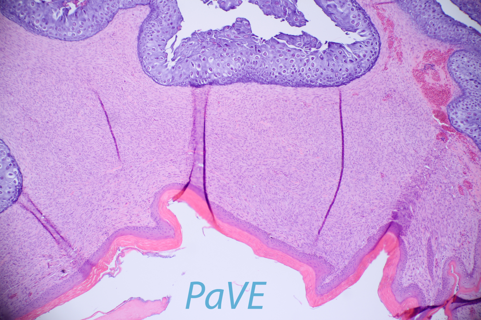
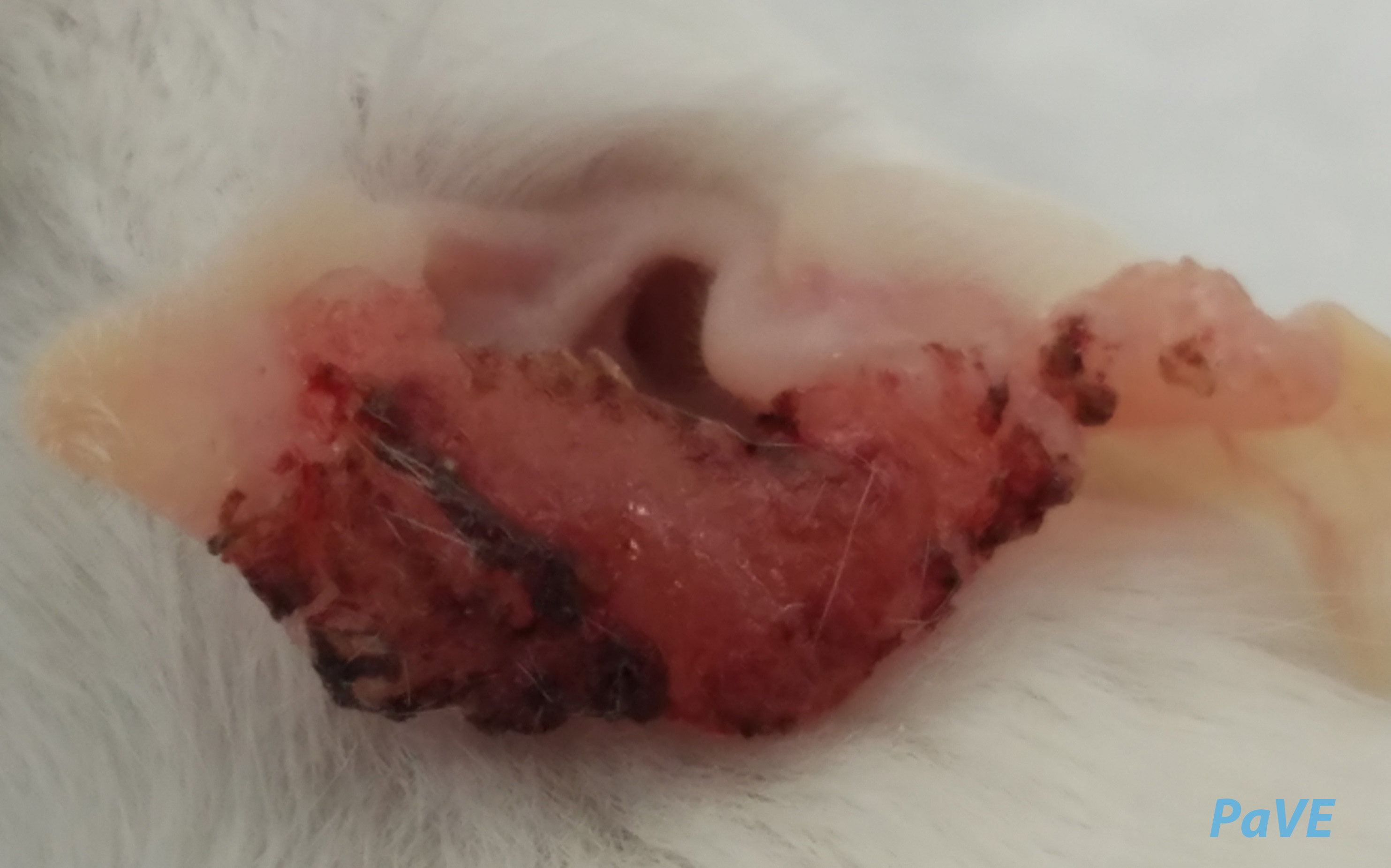
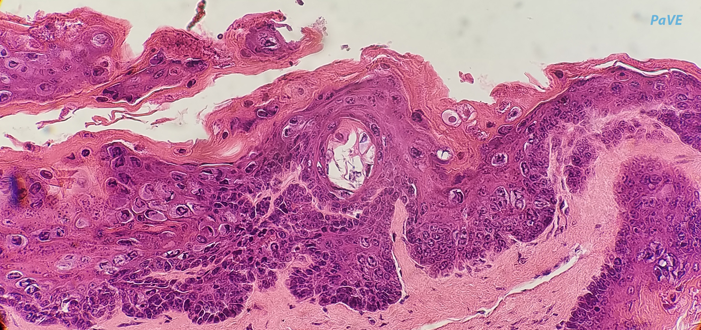
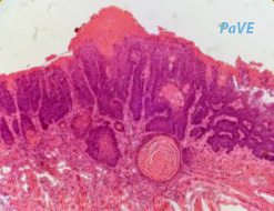
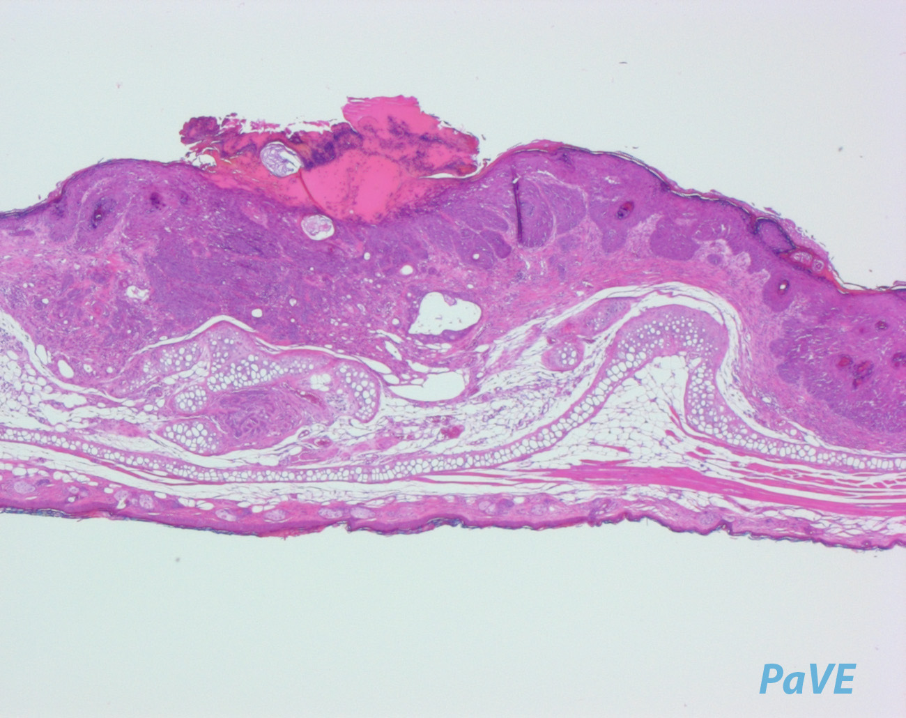
H&E section of a bovine fibropapilloma. Viruses in the delta genus, such as BPV1, cause fibropapillomas in ungulates. The virus infects and replicates in dermal fibroblasts causing extensive proliferation and a large fibroma underlying the epithelium. The overlying epithelium shows papillomatosis, acanthosis, koilyocytosis, parakeratosis and hyperkeratosis.
Author: Baker
Credit: Carl Baker. This image is the work of an National Institutes of Health employee, taken or made as part of that person's official duties. As a work of the U.S. federal government, the image is in the public domain.
Penile fibropapilloma/carcinoma. Lesions are due to BPV1
Author: Campo and Jarrett
Credit: Saveria Campo, Glasgow University.
Teat papillomas in a cow. Large front fibropapillomas are caused by BPV-1; small oval fibropapillomas ('rice grain') by BPV-5, and small frond epithelial papillomas by BPV-6
Author: Campo and Jarrett
Credit: Reprinted from Campo, Animal models of papillomavirus pathogenesis, Virus Research\n89, 249-261 (2002) with permission from Elsevier.
Several studies have found an association between the presence of Bovine papillomavirus-1 and 2 and associated viral growth proteins in skin cells with sarcoid formation in horses. Occult (hairless area at left) and nodular (large round bump at right) forms of equine sarcoids.
Author: Malcolm Morely
Credit: Malcolm Morely, GNU Free license, Wikimedia
H&E stain of a bovine fibropapilloma
Author: Pesavento
Credit: Patricia Pesavento, UC Davis School of Veterinary Medicine
H&E stain of a bovine fibropapilloma
Author: Pesavento
Credit: Patricia Pesavento, UC Davis School of Veterinary Medicine
Immunohistochemistry of a bovine fibropapilloma
Author: Pesavento
Credit: Patricia Pesavento, UC Davis School of Veterinary Medicine
In situ hybridization for BPV1 E5 and E6 regions in a bovine fibropapilloma
Author: Pesavento
Credit: Patricia Pesavento, UC Davis School of Veterinary Medicine
H&E stain of equine sarcoid
Author: Pesavento
Credit: Patricia Pesavento, UC Davis School of Veterinary Medicine
In situ hybridization (BPV1) of equine sarcoid
Author: Pesavento
Credit: Patricia Pesavento, UC Davis School of Veterinary Medicine
H&E stain of equine sarcoid
Author: Pesavento
Credit: Patricia Pesavento, UC Davis School of Veterinary Medicine
In situ hybridization (BPV1) of equine sarcoid
Author: Pesavento
Credit: Patricia Pesavento, UC Davis School of Veterinary Medicine
H&E stain of equine sarcoid
Author: Pesavento
Credit: Patricia Pesavento, UC Davis School of Veterinary Medicine
In situ hybridization (BPV1) of equine sarcoid
Author: Pesavento
Credit: Patricia Pesavento, UC Davis School of Veterinary Medicine
H&E stain of equine sarcoid
Author: Pesavento
Credit: Patricia Pesavento, UC Davis School of Veterinary Medicine
In situ hybridization (BPV1) of equine sarcoid
Author: Pesavento
Credit: Patricia Pesavento, UC Davis School of Veterinary Medicine
Equine sarcoid
Author: Pesavento
Credit: Patricia Pesavento, UC Davis School of Veterinary Medicine
Equine sarcoid
Author: Pesavento
Credit: Patricia Pesavento, UC Davis School of Veterinary Medicine
Equine sarcoid
Author: Pesavento
Credit: Patricia Pesavento, UC Davis School of Veterinary Medicine
BPV2
Infection with bovine papillomavirus type 1 (BPV1) or BPV2 induces fibropapillomas in cows and skin sarcoids in horses.
Author: Campo and Jarrett
Credit: Saveria Campo, Glasgow University.
BPV4
Oesophageal papillomas in cattle. These lesions are positive for BPV4
Author: Campo and Jarrett
Credit: Saveria Campo, Glasgow University.
Florid papillomatosis of pharynx caused by infection with BPV4
Author: Campo and Jarrett
Credit: Reprinted from Campo, Animal models of papillomavirus pathogenesis, Virus Research\n89, 249-261 (2002) with permission from Elsevier.
Florid papillomatosis of rumen caused by infection with BPV4
Author: Campo and Jarrett
Credit: Reprinted from Campo, Animal models of papillomavirus pathogenesis, Virus Research\n89, 249-261 (2002) with permission from Elsevier.
Florid papillomatosis of reticulum caused by infection with BPV4
Author: Campo and Jarrett
Credit: Reprinted from Campo, Animal models of papillomavirus pathogenesis, Virus Research\n89, 249-261 (2002) with permission from Elsevier.
BPV4 containing papilloma of the palate
Author: Campo and Jarrett
Credit: Reprinted from Campo, Animal models of papillomavirus pathogenesis, Virus Research\n89, 249-261 (2002) with permission from Elsevier.
Neoplastic lesion in bracken grazing cattle
Author: Campo and Jarrett
Credit: Reprinted from Campo, Animal models of papillomavirus pathogenesis, Virus Research\n89, 249-261 (2002) with permission from Elsevier.
Florid papillomas of the GI tract in cattle grazing on bracken fern. Lesions are caused by BPV4
Author: Campo and Jarrett
Credit: Saveria Campo, Glasgow University.
GI tract cancer in cattle grazing on bracken fern. Lesions are caused by BPV4
Author: Campo and Jarrett
Credit: Saveria Campo, Glasgow University.
Histopathological (H&E) stain of a papilloma caused by BPV4
Author: Campo and Jarrett
Credit: Saveria Campo, Glasgow University.
Histopathological (H&E) stain of a transforming papilloma caused by BPV4
Author: Campo and Jarrett
Credit: Saveria Campo, Glasgow University.
Histopathological (H&E) stain of a carcinoma caused by BPV4
Author: Campo and Jarrett
Credit: Saveria Campo, Glasgow University.
BPV5
Teat papillomas in a cow. Large front fibropapillomas are caused by BPV-1; small oval fibropapillomas ('rice grain') by BPV-5, and small frond epithelial papillomas by BPV-6
Author: Campo and Jarrett
Credit: Reprinted from Campo, Animal models of papillomavirus pathogenesis, Virus Research\n89, 249-261 (2002) with permission from Elsevier.
BPV6
Teat papillomas in a cow. Large front fibropapillomas are caused by BPV-1; small oval fibropapillomas ('rice grain') by BPV-5, and small frond epithelial papillomas by BPV-6
Author: Campo and Jarrett
Credit: Reprinted from Campo, Animal models of papillomavirus pathogenesis, Virus Research\n89, 249-261 (2002) with permission from Elsevier.
CdPV1
A dromedary calf with cauliflower-like papillomas on the muzzle and lower jaw. CdPV1 was isolated from these lesions. Photograph was takein in August 2009 in Al-Qutaynah locality (South of Khartoum) by Ahmed K. Elfadl, Pathology dept., Faculty of Veterinary Medicine, University of Khartoum, Sudan
Author: Forslund
Credit: Ola Forslund, Malmo University
A dromedary calf with multiple papillomas on the upper lip. Photograph was takein in July 2011 in Al-Showak locality (East Sudan) by Ahmed K. Elfadl, Pathology dept., Faculty of Veterinary
Author: Forslund
Credit: Ola Forslund, Malmo University
Histopathological section (H&E) of a camel fibropapilloma. The lesions shows hyperkeratosis, acanthosis and dermal fibrosis
Author: Forslund
Credit: Ola Forslund, Malmo University
ChPV1
ChPV1 was isolated from the health skin of a goat.
The papillomavirus manifests through irregular tumors (typically in shapes resembling cauliflower) and may occur in the mouth or on other areas of the dog's skin. The most common areas affected by the tumors are the muzzle and the lips, but the tumors may grow inside the dog's mouth as well.
Author: Mills
Credit: Joel Mills, GNU Free license, Wikimedia
The papillomavirus manifests through irregular tumors (typically in shapes resembling cauliflower) and may occur in the mouth or on other areas of the dog's skin. The most common areas affected by the tumors are the muzzle and the lips, but the tumors may grow inside the dog's mouth as well.
Author: Yuan and Schlegel, Georgetown
Credit: Yuan and Schlegel, Georgetown University
CPV1 lesions are usually cleared by the host immune system. However, in some cases infection can results in non-regressive oral warts
Author: Yuan and Schlegel, Georgetown University
Credit: Yuan and Schlegel, Georgetown University
Histopathological section of CPV1 lesion (H&E). 10x magnification
Author: Yuan and Schlegel, Georgetown University
Credit: Yuan and Schlegel, Georgetown University
Histopathological section of CPV1 lesion (H&E). 40x magnification
Author: Yuan and Schlegel, Georgetown University
Credit: Yuan and Schlegel, Georgetown University
Photomicrograph of tissue section stained immunohistochemically. 10x magnification
Author: Yuan and Schlegel, Georgetown University
Credit: Yuan and Schlegel, Georgetown University
Photomicrograph of tissue section stained immunohistochemically. 40x magnification
Author: Yuan and Schlegel, Georgetown University
Credit: Yuan and Schlegel, Georgetown University
CPV2
Paw lesion caused by CPV2
Author: Yuan and Schlegel, Georgetown University
Credit: Yuan and Schlegel, Georgetown University
Paw lesion caused by CPV2
Author: Yuan and Schlegel, Georgetown University
Credit: Yuan and Schlegel, Georgetown University
Histopathological section of CPV1 lesion (H&E). 10x magnification
Author: Yuan and Schlegel, Georgetown University
Credit: Yuan and Schlegel, Georgetown University
Histopathological section of CPV1 lesion (H&E). 40x magnification
Author: Yuan and Schlegel, Georgetown University
Credit: Yuan and Schlegel, Georgetown University
Photomicrograph of tissue section stained immunohistochemically. 10x magnification
Author: Yuan and Schlegel, Georgetown University
Credit: Yuan and Schlegel, Georgetown University
Photomicrograph of tissue section stained immunohistochemically. 40x magnification
Author: Yuan and Schlegel, Georgetown University
Credit: Yuan and Schlegel, Georgetown University
FcPV1
Papillomatous lesions on the foot of a wildChaffinch. Picture was taken in Ripon, UK. Lesion is likely due to FcPV1
Author: Coze
Credit: Yvonne Elvin and British Trust for Ornithology
Papillomatous lesions on the foot of a wild Chaffinch. Picture was taken in Ripon, UK. Lesion is likely due to FcPV1
Author: Coze
Credit: Yvonne Elvin and British Trust for Ornithology
Papillomatous lesions on the foot of a wild Chaffinch. Picture was taken in Ripon, UK. Lesion is likely due to FcPV1
Author: Coze
Credit: Yvonne Elvin and British Trust for Ornithology
Papillomatous lesions on the foot of a wild Chaffinch. Picture was taken in Ripon, UK. Lesion is likely due to FcPV1
Author: Coze
Credit: Yvonne Elvin and British Trust for Ornithology
Papillomatous lesions on the foot of a wild Chaffinch. Picture was taken in Ripon, UK. Lesion is likely due to FcPV1
Author: Coze
Credit: With permission from www.cycoze.com
FlPV1
FlPV1 was isolated from the healthy skin of yellow necked spur fowl
Author: Stolz
Credit: Gary M. Stolz. This image is the work of a U.S. Fish and Wildlife Service employee, taken or made as part of that person's official duties. As a work of the U.S. federal government, the image is in the public domain.
HPV
Section of exophytic wart showing hyperkeratosis of upper layers of epidermis and hypertrophy of basal layers
Author: Cubie
Credit: Heather Cubie, Edinburgh University
Florid persistent hand warts on the hand of a chicke fascia handler
Author: Cubie
Credit: Heather Cubie, Edinburgh University
Extensive atypical plane warts found in patient with Epidemodysplacia verruciformis
Author: Cubie
Credit: Heather Cubie, Edinburgh University
Histology of an Epidemodysplacia verruciformis-like lesion showing characteristic large clear cells in spinous and granular layers
Author: Cubie
Credit: Heather Cubie, Edinburgh University
Mosaic plantar wart in liver transplant recipient
Author: Cubie
Credit: Heather Cubie, Edinburgh University
Renal allograft recipient 1960s treated with high dose steroids + azathioprine
Author: Cubie
Credit: Heather Cubie, Edinburgh University
Extensive persistent warts from childhood with severe familial CD4 lymphopaenia
Author: Cubie
Credit: Heather Cubie, Edinburgh University
Recalcitrant mosaic warts on thumb of young woman after chemotherapy for Hodgkin’s disease
Author: Cubie
Credit: Heather Cubie, Edinburgh University
Extensive Flat warts on shoulder of HIV infected patient
Author: Cubie
Credit: Heather Cubie, Edinburgh University
Anogenital warts in adult
Author: Cubie
Credit: Heather Cubie, Edinburgh University
Anogenital warts on labia of child
Author: Cubie
Credit: Heather Cubie, Edinburgh University
Single genital wart on buttock of child
Author: Cubie
Credit: Heather Cubie, Edinburgh University
Extensive perianal warts in renal allograft recipient
Author: Cubie
Credit: Heather Cubie, Edinburgh University
Extensive perianal warts in child
Author: Cubie
Credit: Heather Cubie, Edinburgh University
Colposcopic appearance of flat warts on cervix (CIN 3)
Author: Cubie
Credit: Heather Cubie, Edinburgh University
Anal Intraepithelial Neoplasia (AIN) in an HIV+ individual
Author: Cubie
Credit: Heather Cubie, Edinburgh University
Early stromal invasion in a CIN3 lesion
Author: Cubie
Credit: Heather Cubie and Alistair Williams, Edinburgh University
Cytological image of HPV associated pre-cancer
Author: Cubie
Credit: Heather Cubie, Edinburgh University
Cytological image of HPV associated pre-cancer
Author: Cubie
Credit: Heather Cubie, Edinburgh University
Cytological image of HPV associated pre-cancer
Author: Cubie
Credit: Heather Cubie, Edinburgh University
Cytological image of HPV associated pre-cancer
Author: Cubie
Credit: Heather Cubie, Edinburgh University
Cytological image of HPV associated pre-cancer
Author: Cubie
Credit: Heather Cubie, Edinburgh University
Histological image of HPV associated pre-cancer
Author: Cubie
Credit: Heather Cubie and Alistair Williams, Edinburgh University
Histological image of HPV associated pre-cancer
Author: Cubie
Credit: Heather Cubie and Alistair Williams, Edinburgh University
Histological image of HPV associated pre-cancer
Author: Cubie
Credit: Heather Cubie and Alistair Williams, Edinburgh University
Histological image of HPV associated pre-cancer
Author: Cubie
Credit: Heather Cubie and Alistair Williams, Edinburgh University
Histological image of HPV associated pre-cancer
Author: Cubie
Credit: Heather Cubie and Alistair Williams, Edinburgh University
Oral lesions in patient with AIDS
Author: Cubie
Credit: Heather Cubie, Edinburgh University
Warty lesion resembling condyloma on base of the tongue
Author: McMillan and Cubie
Credit: Heather Cubie and Alistair Williams, Edinburgh University. By permission of Oxford University Press VIRAL WARTS: BIOLOGY AND TREATMENT by Mary H. Bunney, Claire Benton & Heather A. Cubie (1992).
Condylomata acuminate on glans penis and foreskin
Author: McMillan and Cubie
Credit: Heather Cubie and Alistair Williams, Edinburgh University. By permission of Oxford University Press VIRAL WARTS: BIOLOGY AND TREATMENT by Mary H. Bunney, Claire Benton & Heather A. Cubie (1992).
Thrombosed capillaries in plantar wart due to spontaneous resolution
Author: Cubie
Credit: Heather Cubie, Edinburgh University. By permission of Oxford University Press VIRAL WARTS: BIOLOGY AND TREATMENT by Mary H. Bunney, Claire Benton & Heather A. Cubie (1992).
Mosaic plantar wart in liver transplant recipient
Author: Cubie
Credit: Heather Cubie, Edinburgh University
Mosaic plantar wart in liver transplant recipient
Author: Cubie
Credit: Heather Cubie, Edinburgh University
HPV1
Electron micrograph of virus particles in crystalline array in nuclei of granulocytic and superfical layers of HPV1 associated plantar wart
Author: Cubie
Credit: Heather Cubie, Edinburgh University.
Deep plantar warts caused by HPV1
Author: Cubie
Credit: Heather Cubie, Edinburgh University
Deep plantar wart caused by HPV1
Author: Cubie
Credit: Heather Cubie, Edinburgh University
HPV16
Section of cervix showing CIN2 resulting from HPV16 infection. The cervix shows moderate dysplasia with dysplastic cells extending through the middle third of the epithelium.
Author: McBride
Credit: Alison McBride. This image is the work of an National Institutes of Health employee, taken or made as part of that person's official duties. As a work of the U.S. federal government, the image is in the public domain.
In situ squamous cell carcinoma, progressing to invasive disease - such lesions can be associated with HPV16
Author: Cubie
Credit: Heather Cubie, Edinburgh University
HPV2
Multiple common warts across back of hand
Author: Cubie
Credit: Heather Cubie, Edinburgh University
Periungual warts
Author: Cubie
Credit: Heather Cubie, Edinburgh University
Florid hand warts in patient with some immunosuppression
Author: Cubie
Credit: Heather Cubie, Edinburgh University
Mosaic plantar wart caused by HPV2
Author: Cubie
Credit: Heather Cubie, Edinburgh University
Filiform common beard warts
Author: Cubie
Credit: Heather Cubie, Edinburgh University
Common wart arising at site of nose piercing
Author: Cubie
Credit: Heather Cubie, Edinburgh University
HPV3
Plane warts on face and in linear arrangement below lip arising from a scratch (knonw as the Koebner phenomenon)
Author: Cubie
Credit: Heather Cubie, Edinburgh University
Koebner phenomenon demonstrated by arrow along scratch line
Author: Cubie
Credit: Heather Cubie, Edinburgh University
HPV5
Epidemodysplacia verruciformis like plaque in renal allograft recipient (these lesions are associated with HPV5/HPV8 infections)
Author: Cubie
Credit: Heather Cubie, Edinburgh University
Epidemodysplacia verruciformis like plaque in renal allograft recipient (these lesions are associated with HPV5/HPV8 infections)
Author: Cubie
Credit: Heather Cubie, Edinburgh University
HPV7
Florid persistent hand warts found on the hands of a fish handler (HPV7 was detected in these lesions)
Author: Cubie
Credit: Heather Cubie, Edinburgh University
HPV8
Epidemodysplacia verruciformis like plaque in renal allograft recipient (these lesions are associated with HPV5/HPV8 infections)
Author: Cubie
Credit: Heather Cubie, Edinburgh University
Epidemodysplacia verruciformis like plaque in renal allograft recipient (these lesions are associated with HPV5/HPV8 infections)
Author: Cubie
Credit: Heather Cubie, Edinburgh University
RaPV1
Photograph of wing membrane showing raised, irregular skin mass. This is characteristic of the basosquamous carcinoma observed in this animal (Bar=1cm)
Author: Van Ranst
Credit: Reprinted from Journal of Zoo and Wildlife Medicine 37:193-196 (2006 )\n with Permission from American Association of Zoo Veterinarians.
(a) H&E stain of skin mass. Biopt was taken from the side of the face of the animal. Neoplastic appearing epithelial cells extend into the underlying dermis. These cells often surround dilated hair follicles. Bar=25 um. (b)H&E stain of skin mass. Biopt was taken from the side of the face of the animal. Basilar epithelial cells are invading the underlying dermis. Bar=200 um. (c) H&E stain of skin mass. Biopt was taken from the left wing of the animal. Neoplastic cells can be surrounding cells undergoing squamous differentiation.
Author: Van Ranst
Credit: Reprinted from Journal of Zoo and Wildlife Medicine 37:193-196 (2006 )\n with Permission from American Association of Zoo Veterinarians.
SfPV1
The picture below was sent to Dan Japuntich by Grant VanGilder of Mankato, MN. It shows a severe infection with the Shope papillomavirus, although Grant says, “He is still alive and kicking and is the talk of the neighborhood.” (As of 5/19/03) Thanks, Grant!
Author: Vangilder
Credit: Grant VanGilder, MN, USA
Photograph of gross specimen as it was presented to the Diagnostic Laboratory at Colorado State University
Author: Weiner
Credit: Reproduced from J VET Diagn Invest 2010 22: 451. Permission for further reuse must be requested from SAGE Publications
Photomicrograph of tissue section stained immunohistochemically using antibodies generated against chemically-disrupted bovine papilloma virus type I. Positive staining by Nova Red chromogen is indicated by the bright red intranuclear inclusion material
Author: Weiner
Credit: Reproduced from J VET Diagn Invest 2010 22: 451. Permission for further reuse must be requested from SAGE Publications
A- The chromatin in the nuclei of the papilloma cells is marginated. Aggregates of chromatin clumps are seen in clear nucleus filled with electron-dense virions. The nucleolus has reticular pattern nucleolonema and may be decreased in size. (Scale bar: 1µm); B- Higher power image of the framed area in Figure 2A. Note the nuclear pore (arrow) and papilloma virions (open arrows). (Scale bar: 100 nm); C- The luminal contents of a blood vessel include a Langerhans cell (LC), which is identified by its characteristic cytoplasmic Birbeck granules, also called Langerhans’ cell granules (arrow). The boxed areas are shown at a higher magnification in panels D and E. (Scale bar: 2 µm); D- Higher power image of LC Birbeck granules (arrow). (Scale bar: 200 nm); E- Within the Langerhans cell are many papillomavirus virions (open arrows). This demonstrates the direct involvement of LCs in immune trafficking during papillomavirus infection and corroborates previous reports that LCs enhance cell-mediated immunity during papillomavirus infection. (Scale bar: 200 nm)
Author: Weiner
Credit: Reproduced from J VET Diagn Invest 2010 22: 451. Permission for further reuse must be requested from SAGE Publications
Langerhans cells are within the lumen of blood vessels amidst erythrocytes, and endothelial cells lining vessel walls were evident. (Scale bar: 2 um)
Author: Weiner
Credit: Reproduced from J VET Diagn Invest 2010 22: 451. Permission for further reuse must be requested from SAGE Publications
This is a specimen captured in Topeka Kansas, and it is held at the University of Kansas Natural History Museum and Biodiversity Research Center. Picture by Heather York
Author: York
Credit: Heather York
UuPV1
Oral papillomatous lesions in a snow leopard. The pale nodules on the bottom of the tongue are papillomas and are indicated by the black circle. The papillomatous lesions are arranged in a ring. This circular arrangement is likely the result of an earlier biopsy in this animal. During the biopsy, viral particles seeded the surrounding skin and caused additional papillomas to form in a circle around the original site of ablation.
Author: Mitsouras
Credit: Reprinted from BMC Veterinary Research 7:38 (2011).This article is distributed under the terms of the Creative Commons Attribution License, which permits unrestricted use, distribution, and reproduction in any medium, provided the original work is properly cited. \n\n
EaPV1
Asinara white donkey: macroscopic features of lesions. On general physical examination, no abnormalities were detected. On dermatological examination the external aspect of both ear pinna were characterized by the presence of white/brownish adherent scales, alopecia end eritema. A few small multifocal areas of dark pigmented skin were also detected. Similar but milder lesions were also detected on the dorsal aspect of the nose and in the periocular lesions.
Author: Alberti
Credit: Dr. Alberto Alberti, University of Sassari
Asinara white donkey: microscopic features of lesions. Epidermis is mildly hyperplastic and shows multifocal vacuolation of the basal layer.
Author: Alberti
Credit: Alberto Alberti, University of Sassari
Asinara white donkey: microscopic features of lesions. Epidermis is mildly hyperplastic and shows multifocal vacuolation of the basal layer.
Author: Alberti
Credit: Alberto Alberti, University of Sassari
HPV14
Clinical picture of an HPV14-positive wart-like lesion from an Epidermodysplasia verruciformis patient.
Author: Cinzia Borgogna and Marisa Gariglio
Credit: Marisa Gariglio, University of Piedmont Region, Novara-Italy
Histology of an HPV14-positive wart-like lesion from an Epidermodysplasia verruciformis patient showing characteristic large clear cells in spinous and granular layers
Author: Cinzia Borgogna and Marisa Gariglio
Credit: Marisa Gariglio, University of Piedmont Region, Novara-Italy
Detection of the viral protein E4 by immunofluorescence with a rabbit polyclonal anti-E4 antibody in an HPV14-positive wart-like lesion from an Epidermodysplasia verruciformis patient showing cytoplasmic staining in spinous and granular layers. The section is counterstained with DAPI (blue) to visualize cell nuclei.
Author: Cinzia Borgogna, John Doorbar and Marisa Gariglio
Credit: Marisa Gariglio, University of Piedmont Region, Novara-Italy
HPV24
Clinical picture of an HPV24-positive wart-like lesion from a patient with a primary T-cell immunodeficiency
Author: Cinzia Borgogna and Marisa Gariglio
Credit: Marisa Gariglio, University of Piedmont Region, Novara-Italy
Histology of an HPV24-positive wart-like lesion from a patient with a primary T-cell immunodeficiency showing characteristic large clear cells in spinous and granular layers
Author: Cinzia Borgogna and Marisa Gariglio
Credit: Marisa Gariglio, University of Piedmont Region, Novara-Italy
Detection of the viral protein E4 by immunofluorescence with a rabbit polyclonal anti-E4 antibody in an HPV24-positive wart-like lesion from a patient with a primary T-cell immunodeficiency showing cytoplasmic staining in spinous and granular layers. The section is counterstained with DAPI (blue) to visualize cell nuclei.
Author: Cinzia Borgogna, John Doorbar and Marisa Gariglio
Credit: Marisa Gariglio, University of Piedmont Region, Novara-Italy
OaPV3
Squamous cell carcinoma in the nasal region of a sarda sheep infected with OaPV3
Author: Alberti
Credit: Alberto Alberti, University of Sassari
Squamous cell carcinoma in the nasal region of a sarda sheep infected with OaPV3
Author: Alberti
Credit: Alberto Alberti, University of Sassari
CPV
Immunohistochemistry of canine inverted papilloma
Author: Pesavento
Credit: Patricia Pesavento, UC Davis School of Veterinary Medicine
Immunohistochemistry of canine inverted papilloma
Author: Pesavento
Credit: Patricia Pesavento, UC Davis School of Veterinary Medicine
Immunohistochemistry of canine inverted papilloma
Author: Pesavento
Credit: Patricia Pesavento, UC Davis School of Veterinary Medicine
H&E stain of a canine inverted papilloma
Author: Pesavento
Credit: Patricia Pesavento, UC Davis School of Veterinary Medicine
H&E stain of a canine inverted papilloma
Author: Pesavento
Credit: Patricia Pesavento, UC Davis School of Veterinary Medicine
H&E stain of a canine inverted papilloma
Author: Pesavento
Credit: Patricia Pesavento, UC Davis School of Veterinary Medicine
Canine oral papillomatosis
Author: Pesavento
Credit: Patricia Pesavento, UC Davis School of Veterinary Medicine
Oral papillomatous lesions in a coyote
Author: Keel
Credit: Kevin Keel, UC Davis School of Veterinary Medicine
Oral papillomatous lesions in a coyote
Author: Keel
Credit: Kevin Keel, UC Davis School of Veterinary Medicine
Oral papillomatous lesions in a coyote
Author: Keel
Credit: Kevin Keel, UC Davis School of Veterinary Medicine
EPV2
Equine epithelial hyperplasia and dysplasia
Author: Pesavento
Credit: Patricia Pesavento, UC Davis School of Veterinary Medicine
Equine epithelial hyperplasia and dysplasia
Author: Pesavento
Credit: Patricia Pesavento, UC Davis School of Veterinary Medicine
Equine hyperplasia and dysplasia: in situ hybridization
Author: Pesavento
Credit: Patricia Pesavento, UC Davis School of Veterinary Medicine
Equine hyperplasia and dysplasia: in situ hybridization
Author: Pesavento
Credit: Patricia Pesavento, UC Davis School of Veterinary Medicine
H&E stain of equine genital squamous cell carcinoma
Author: Pesavento
Credit: Patricia Pesavento, UC Davis School of Veterinary Medicine
In situ hybridization of equine genital squamous cell carcinoma
Author: Pesavento
Credit: Patricia Pesavento, UC Davis School of Veterinary Medicine
H&E stain of metastatic equine genital squamous cell carcinoma
Author: Pesavento
Credit: Patricia Pesavento, UC Davis School of Veterinary Medicine
H&E stain of metastatic equine genital squamous cell carcinoma
Author: Pesavento
Credit: Patricia Pesavento, UC Davis School of Veterinary Medicine
EPV
Equine aural plaque, presumed EPV
Author: Pesavento
Credit: Patricia Pesavento, UC Davis School of Veterinary Medicine
FcaPV
Feline papilloma
Author: Pesavento
Credit: Patricia Pesavento, UC Davis School of Veterinary Medicine
Feline papilloma
Author: Pesavento
Credit: Patricia Pesavento, UC Davis School of Veterinary Medicine